- Published on
De novo design of allosterically controlled enzymes
- Authors

- Name
- Vishal V
- @VishalVignesh_
Notes
Abstract
cellular processes from signaling to metabolic control (1)
denoising diffusion models enable the design of compact de novo enzymes whose activity can be allosterically activated or repressed by designed protein effectors (1)
Intro
Cell signaling cascades 1, metabolic pathways2,3, and motor proteins 4,5 often rely on coupling between enzymatic activity and protein-ligand binding at sites distal to the active site (1)
Distal as in place elsewhere from the active site.
Some native allosteric enzymes:
Cell-signalling cascade: chain reaction that lets a cell detect input and output some action
Metabolic pathways: sequence of chemical reactions where product of one reaction becomes input of another
Motor proteins: tiny motors that use ATP to create motion. Ex: kinesin walking on a microtubule using ATP carrying transport vesicle
sidechain communication networks relay changes from an allosteric binding site to the enzyme active site (1)
A mechanistic model.
allosteric effectors act more broadly to shift the equilibria between well-defined distinct states (1)
A chemical equilibrium model.
to design allosteric systems in synthetic biology have generally relied on natural enzymes engineered with topological rearrangements11,12, insertions/fusions13, and split chains 14,15 to couple a protein-ligand binding event to enzymatic activity (1)
Before De Novo approach.
such domain rearrangements do not structurally recapitulate the allostery in many naturally evolved enzyme folds, which are often compact single-domain proteins (2)
Prior approaches can't generally achieve the single-domain-ness,
That is,
simply one structural unit instead of seperate domains for binding, calatysis and regualtion.
de novo design of allosteric enzymes remains an outstanding challenge (2)
protein effector. (2)
Effector: molecule that binds a protein and changes its activity.
aimed to more closely emulate natural allosteric control, where a compact structure switches between subtly different conformations that differ in catalytic activity (2)
an active state, where the catalytic residues are optimally positioned to mediate the reaction, (2)
inactive state, where these residues deviate from this catalytically optimal geometry (2)
faster switching between states, as the energy barriers to conversion between very closely related states (2)
Small energy barrier in general.
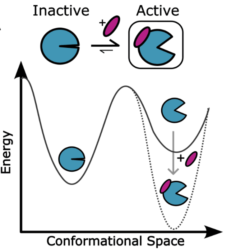
Allosteric Activator.
Effector to bind preferentially to active state.
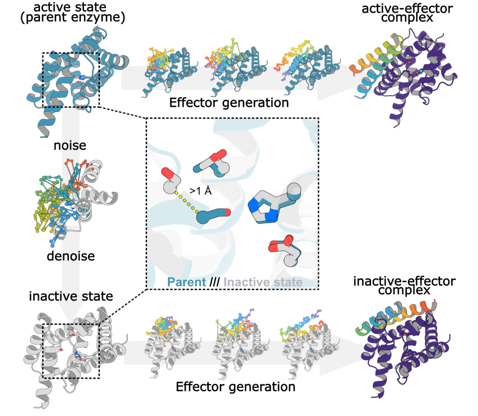
Domain 2 (Ser-Thr) is partially diffused.
Alpha-C is displaced by 1 Angstorm
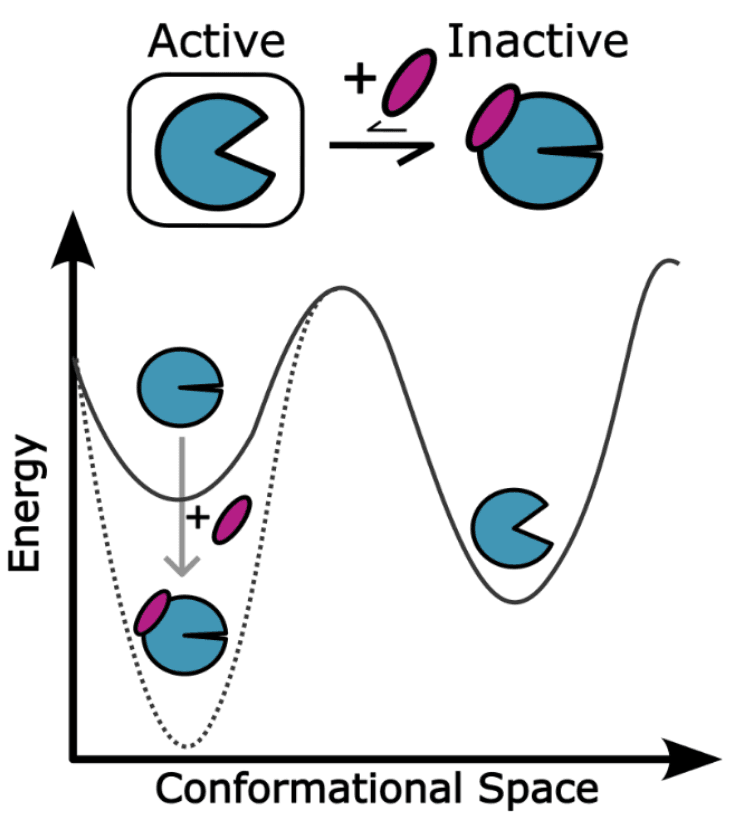
Allosteric Inhibitor.
Effector to bind preferentially to inactive state.
partially diffused (downward arrow) by noising and denoising a portion of the protein (colored in the middle panel at the left, the gray portion is kept fixed) (4)
Partial RFdiffusion: To generate inactive state.
RFdiffusion is then used to generate binders to either the active state (top) in the allosteric activator case, or the inactive state (allosteric inhibitor case) (4)
RFdiffusion: To generate binders for active and inactive states.
Sequence design using tiedMPNN is used to favor the inactive state in the absence of effector in the activator case, and the active state in the absence of effector in the inhibitor case (4)
For allosteric activator:
G(E_I) < G(E_A)
But not specified here.
Design approach
Starting from an active designed enzyme (Fig 1A, “Active”), we generate an alternative inactive state by partial RFdiffusion (Fig 1B) (4)
Typo: (Fig 1B, "Active") and (Fig 1B, "Inactive")
1-3Å of random Gaussian noise (4)
Noising.
generate alternative conformations (4)
Denoising.
partial diffusion has had success in identifying subtly modified backbones with increased binding affinity (4)
Emperical observation: Very small changes in confirmation has larger impact on binding energy.
Hence, only a part of the site is noised and denoised, and that is enough.
design allosteric effectors that bind to either the active or inactive state and modulates the energy landscape (4)
allosteric activation systems in which the unbound enzyme has lower energy in the inactive state (Fig 1A, top left), and effector binding favors the active state (4)
allosteric inactivation systems (Fig 1A, top right) where effector binding conversely favors the inactive state (4)
effector-dependent activator design, we used RFDiffusion to generate binders to the active enzyme state (4)
effector-dependent inactivator designs, we used RFDiffusion to generate effectors to inactive enzyme states (4)
tied ProteinMPNN to generate sequences compatible with the backbones of the enzyme–effector complex as well as the isolated enzyme and effector states (4)
de novo serine hydrolase Win1 as a model system as the catalytic mechanism and active-site geometry are well understood (4)
Win1 is used as a model system only.
The sequence is shared between the generated inactive and active states.
So, the sequence isn't same as Win1.
vast majority of partially diffused conformations are likely to have compromised active sites with reduced or ablated catalytic activity (4)
divided the structure into two separate domains: one containing the His–Asp (catalytic dyad) and the other the catalytic Ser and oxyanion Thr (4)
Partially diffuse the Ser-Thr domain.
filtered for Cα displacements greater than 1 Å from the starting active-state structure (4)
Designs predicted by AlphaFold2 to adopt the target active and inactive conformations in the presence and absence of effector, respectively, were selected for experimental characterization (4)
AF2 should predict E_AP_A complex to show active state and E_I is the enzyme alone which shows inactive state.
Characterization of allosteric activators
generated inactive win1 conformations by partial diffusion in the absence of substrate (5)
designed small proteins that bind to and stabilize the active state of win1 (5)
Sixty-one such designed enzyme-effector pairs that passed AF2 filters were selected for experimental characterization (5)
expressed in E. coli (5)
Both enzyme and effector separately.
activity was measured in cell lysates using the fluorogenic substrate 4-methylumbelliferone acetate (4Mu-Ac) (5)
Experimental Assay:
Cell lysate: Make living cells produce the design protein, then break the cells open.
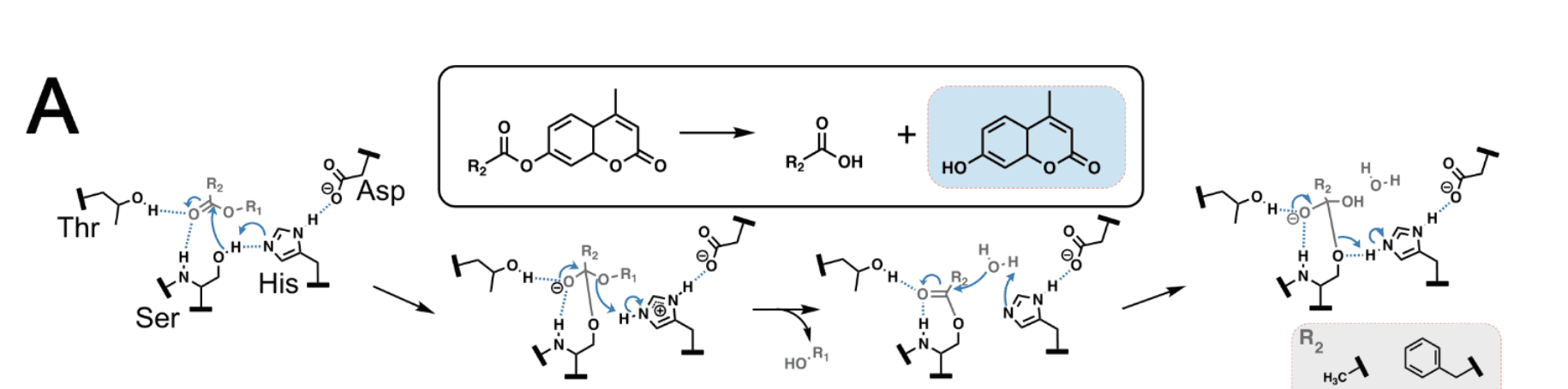
4Mu-Ac + H2O -> 4Mu (fluorescent)+ Ac
Catalyst is serine hydrolase.
identified two effector-enzyme pairs, Janus1 and Janus2 (5)
Both of their active state geometries are similar.
addition of effector cell lysate substantially increased formation of the fluorescent product, 4Mu (5)
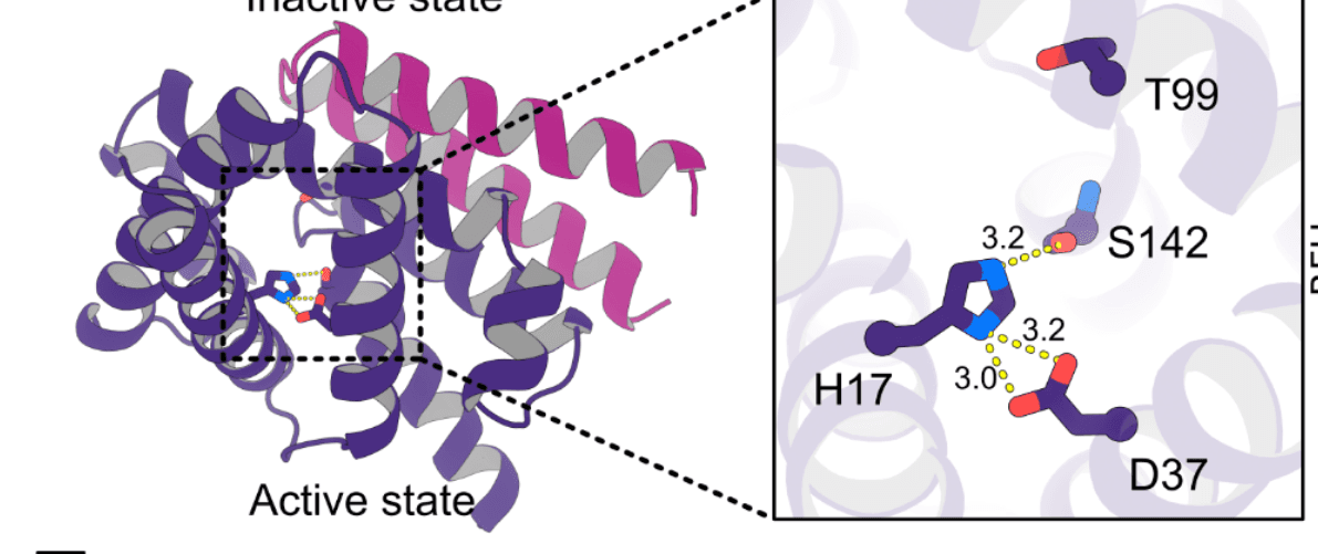
both pairs, there is substantial predicted conformational remodeling of the designed enzyme in the inactive and effector-bound complexes with Cα RMSDs of 8.3 Å and 3.80 Å for Janus1 and Janus2 respectively (5)
nactive state, the catalytic residues Ser142, His17, Asp37, and Thr99 have a disrupted geometry incompatible with efficient catalysis (5)
effector binding, both designs transition to an active-like catalytic arrangement (5)
mutational and kinetic analyses of the purified effectors and enzymes (5)
Mutational Analysis: change / knockout specific residues and see change in catalytic activity
Kinetic Analysis: Measure reaction rates quantitatively
Active site residue knockouts had activity reduced to background levels or below the detection limit (Supplementary Figure 1), supporting catalysis through the expected serine/cysteine hydrolase machinery, as for the parent enzyme (5)
Mutational Analysis.
To characterize the kinetics in more detail, steady-state kinetic measurements were performed in the absence and presence of effector at a 1:2 enzyme:effector ratio (5)
Kinetic Analysis.
Janus1, effector binding increased the turnover rate from kcat = 0.0033 ± 0.00010 s−1 to 0.017 ± 0.0003 s−1, an approximately 5-fold increase (5)
Janus2, kcat increased from 0.00050 ± 0.00005 s−1 in the absence of effector to 0.0013 ± 0.00003 s−1 in the presence of effector, corresponding to a 2.6-fold enhancement (5)
Effector binding also altered substrate affinity in Janus1: Km increased 4.3-fold, from 8.1 ± 1.0 μM in the apo state to 35 ± 2 μM in complex with Janus1_b (5)
Increased K_m results in worse substrate affinity.
Low K_m implies the enzyme works well at low [S]
In contrast, the Km of Janus2 was unchanged within error (4 ± 2 μM apo vs. 2.5 ± 0.3 μM bound (5)
Reduced K_m but insignificant.
2x molar ratio of effector was added to an ongoing enzymatic reaction (5)
both Janus1 and Janus2, addition of effector increased catalytic activity to the levels observed in the pre-incubation experiment (5)
effector on-rate is fast relative to the mixing and measurement dead time (~21 seconds (5)
To test the specificity of each effector for its cognate enzyme, we incubated Janus1 and Janus2 with Janus2_b and Janus1_b, respectively. Both enzymes demonstrate specificity to their respective effector, with no rate enhancement when incubated with the non-cognate effector (Supplementary Figure 2) (5)
Janus1, Active state.
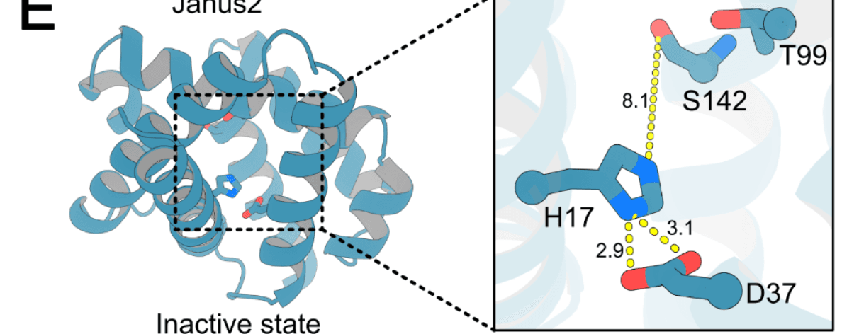
Janus2, Inactive state.
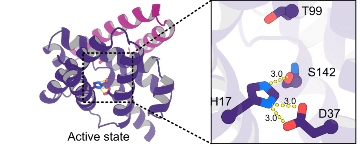
Janus2, Active state.
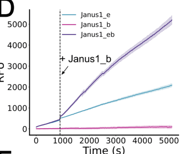
Relative Fluorescence Units.
Slope of this graph is a proxy for enzyme reaction rate.
fluorescent group, R1 in blue, from the leaving group R2 (acetate or phenylacetate) (7)
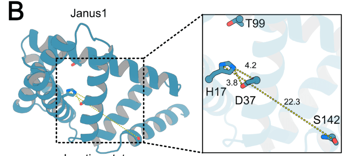
Janus1, Inactive state.
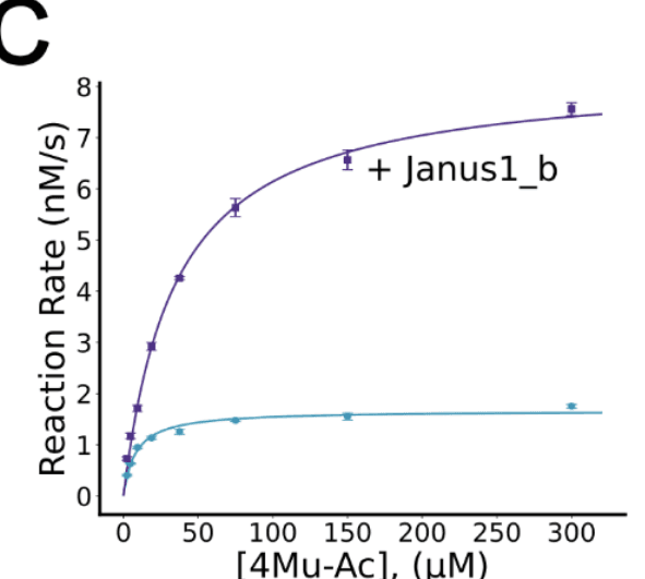
Michaelis-Menten Kinetics.
Michaelis-Menten kinetics of Janus1 comparing unbound enzyme and enzyme with saturating effector (8)
Addition of saturating Janus1_b effector rapidly accelerates catalysis (8)
RFU: Relative Fluorescence Units.
Characterization of allosteric inhibitors
designs off of designed serine hydrolases, win1_b1 16 and win1_b4 (Supplementary Figure 1), that hydrolyze the larger substrate 4-methylumbelliferone phenylacetate (4Mu-PhAc, Figure 2A) (8)
The system changes from allosteric activator case to allosteric inhibitor case.
coupling in molecular machines like Hsp70 24, in which ATP binding switches Hsp70 into a protein-binding competent state, we reasoned that the bulkier, more hydrophobic leaving group of 4Mu-PhAc would provide more binding energy to drive conformational change (8)
4Mu-PhAc is bulkier than 4Mu-Ac.
del G_bind (S, E) is lower:
when S = 4Mu-PhAc
than when S = 4Mu-Ac.
sought to improve the baseline catalytic activity of the designed enzymes in the unbound form (8)
found that a serine-to-cysteine substitution led to dramatic increases in catalytic efficiency across a variety of designed serine hydrolases (8)
Enzyme changed.
experimentally characterized 24 designed allosteric inhibitor-enzyme pairs using lysate screening with fluorescent 4Mu-PhAc in the absence and presence of the effector (8)
Of these, eight designs retained catalytic activity, and one, Janus3, had a clear reduction of enzymatic activity for both cysteine and serine variants upon addition of effector (8)
Cα RMSDs between states of 1.84 Å (8)
Much lower / subtle than activator Janus1, Janus2.
effector binding, the nucleophilic Cys142 is disrupted and turned outward and away (8)
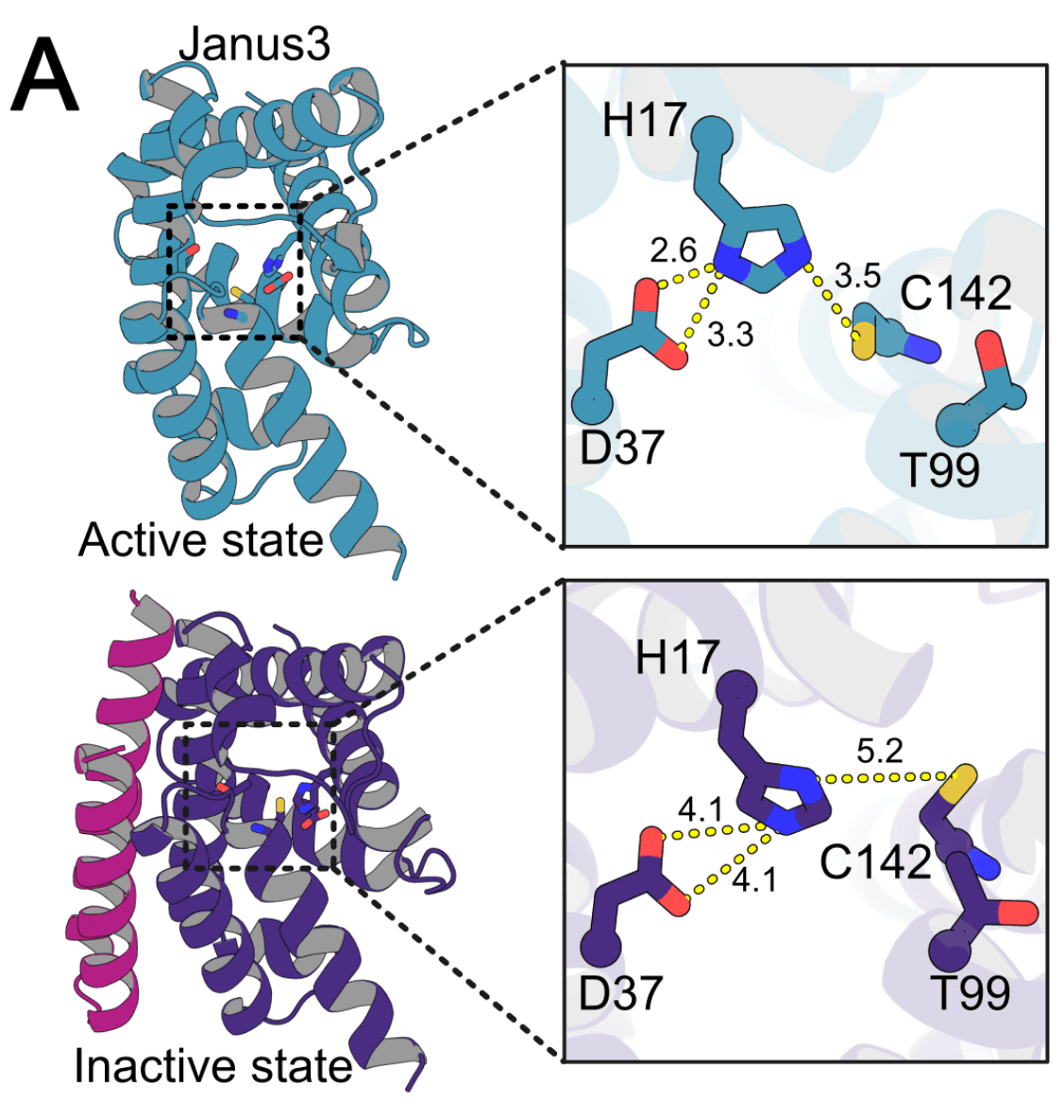
Allosteric Inhibitor.
The primary change is in the catalytic nucleophile residue (9)
Serine to Cystine.
Janus3 enzyme (9)
Janus3_e
effector (Janus3_b) (9)
Binder.
substantial decrease in kcat upon Janus3_b binding (Figure 3B) from .0024 s−1 (unbound) to .0003 s−1 (bound), an 8-fold reduction (Table 1) (9)
no significant difference in KM between the bound and unbound states of Janus3, suggesting that the binding of Janus3_b alters catalytic residue geometry without affecting the substrate binding pocket of Janus3, consistent with the design model (9)
Much cleaner than Janus1 and Janus2.
Mutation of active site residues Cys142, His17, Asp37, and Thr99 to alanine eliminated or reduced activity, confirming that the catalytic mechanism is unchanged (9)
addition of Janus3_b into the Janus3_e and 4Mu-PhAc reaction, the rate decreased to a lower steady state within the dead time of our mixing procedure (9)
Dead time: Experimentally unobservable time, like mixing time so instrument can't measure.
21 secs in the activator case.
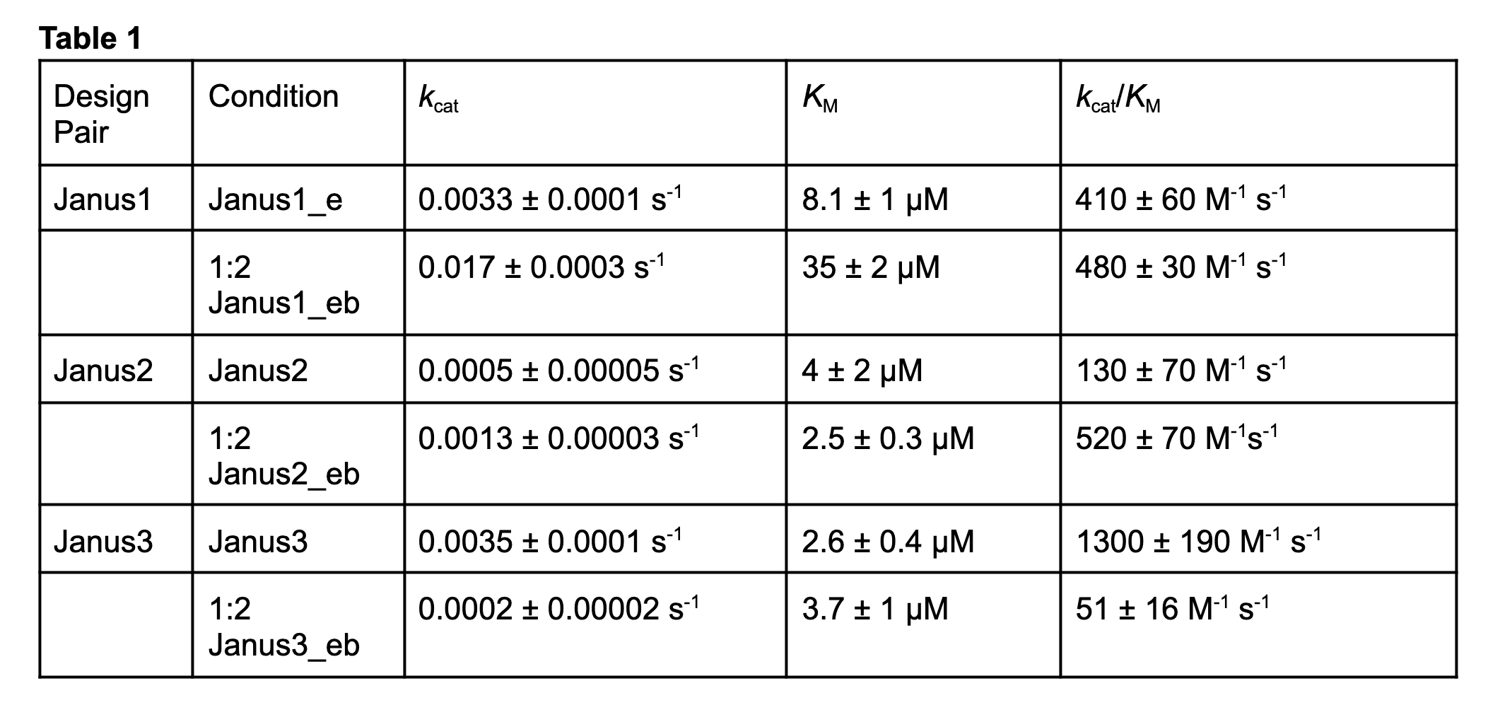
k_cat: Catalytic Rate Constant
K_M: Michaelis Constant
k_cat/K_M: Catalytic Efficiency (Specificity Constant)
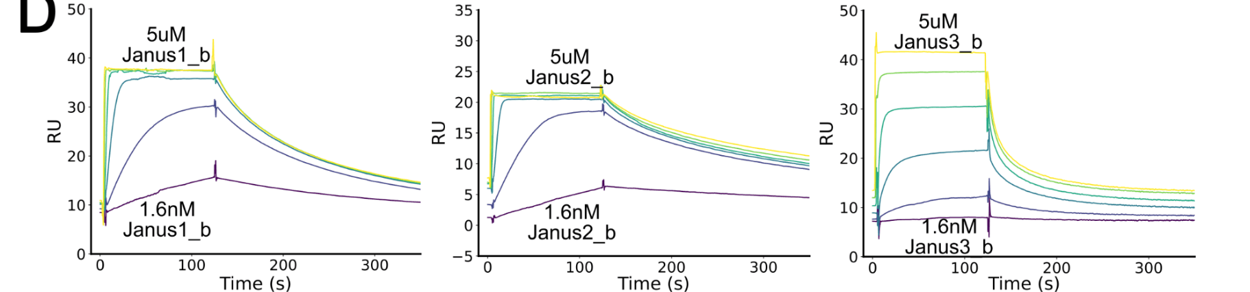
SPR, 5 fold dilutions.
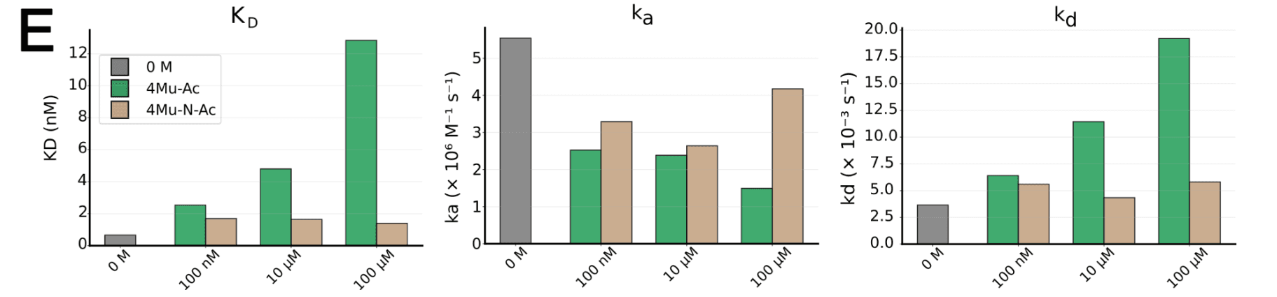
Only Janus1 changed under varying substrate concentration.
K_D = k_d / k_a
4Mu-N-Ac is a non hydrolysable analogue of 4Mu-Ac
SEC traces for the Janus1, Janus2, and Janus3 systems with enzyme alone, effector alone, and enzyme and effector in a 1:1 stoichiometric ratio (11)
SEC: Size Exclusion Chromatography
Inject protein sample into a column packed with porous beads, then continuously pump liquid through it.
Small proteins enter lots of little pores, takes longer route, come out later
Large proteins enter can't enter many pores, take shorter route, come out earlier
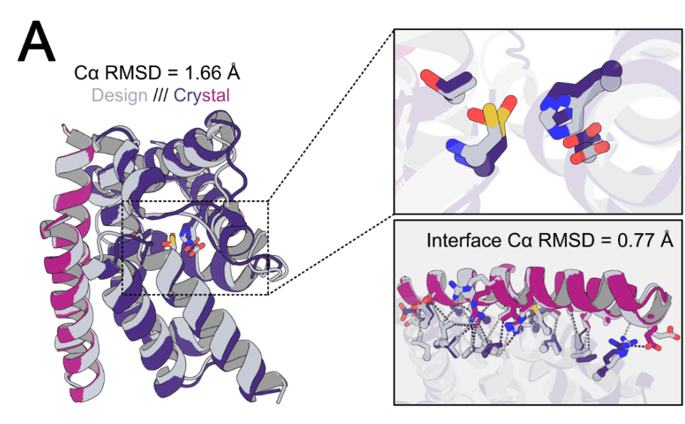
3D design overlaid on the crystal to see if enzyme-effector complex indeed follows the design.
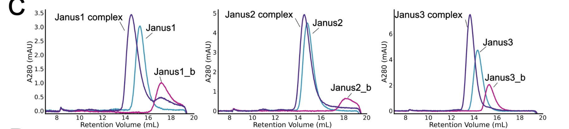
Retention Volume: Amount of liquid has flowed through
usually,
complex > enzyme > binder
A280: Proteins absorb UC at 280nm, because of aromatic amino acids
mAU: milli-Absorbance Units
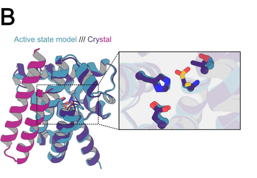
Comparison between the complex (inactive state) vs the unbound active state of the enzyme.
crystal structure of the Janus3 enzyme-effector complex at 1.7 Å resolution (12)
effector binds far from the active site, confirming that the alteration in activity occurs purely due to conformational coupling (rather than steric interference with substrate binding) (12)
catalytic cysteine was oxidized to sulfinic acid, which likely occurred during crystallization or data collection (12)
crystal-inactive state design model Cα RMSD= 1.66 Å, crystal-active state design model Cα RMSD= 1.87 Å (12)
primary differences between the designed active vs inactive states lie in a shift in the placement of the cysteine residue and a rotation and translation of the Thr99 oxyanion stabilizing residue (12)
So the crystal should be similar to the bound state (as intended) but then they also find the active state catalytic site geometry is also very similar. Only a couple of differences:
- shift in cysteine
- rotation and translation of Thr
crystal and the inactive state design model show a 1.2 Å shift of the cysteine backbone Cα relative to the active state model in addition to the rotation of the oxyanion stabilizing oxygen away from the catalytic pocket (12)
Validation that the computational model from active to inactive model state is exhibited by the inactive crystal.
next investigated the effect of substrate on effector binding (12)
- SEC
- SPR
first characterized binding of effector and enzyme in the absence of substrate (12)
By SEC.
Size exclusion chromatography (SEC) of effector alone, enzyme alone, and enzyme and effector together revealed monodisperse 1:1 binding (12)
monodisperse binding: the complex behaves as a uniform species.
E was E B was B EB was EB instead of EB_2, E_2B or any other aggregates
Janus2 enzyme alone appears to be a dimer, but forms a monodisperse complex with Janus2_b with the expected molecular weight (12)
E_2 instead of E for Janus2.
monodisperse complex peak upon addition of effector indicates near stoichiometric binding under these conditions (12)
For 1 E, 1 B binded.
measured the binding affinity of the designed effectors for their cognate enzymes by surface plasmon resonance (SPR) (12)
streptavidin-biotin conjugation (12)
All of the allosteric effectors bind their cognate enzymes with nanomolar to sub-nanomolar affinity: equilibrium dissociation constants (KD) of 2.97 nM for Janus1 (kon = 2.52 × 10⁶ M−1s−1, koff = 7.46 × 10⁻³ s−1), 0.726 nM for Janus2 (kon = 3.49 × 10⁶ M−1s−1, koff = 2.53 × 10⁻³ s−1), and 106 nM for Janus3 (kon = 5.93 × 10⁵ M−1s−1, koff = 6.31 × 10⁻² s−1) (12)
E + B <-> EB
k_on: association rate constant binding rate = k_on[E][B]
k_off: dissociation rate constant unbinding rate = k_off[EB]
K_D = k_off/k_on
Janus3 on-rate is roughly an order of magnitude lower than those of Janus1 and Janus2, suggesting lower occupancy of the inactive binding-competent conformation and higher occupancy of the unbound, active state (12)
This is experimental explanation for the underlying thermodynamics:
They say both active and inactive states exist even when unbounded.
Janus3 takes time to attach with its binder because the Janus3 population has lesser inactive state confirmations to bind to and stabilize.
Janus 1 and Janus 2 has their binder-compatible active state more than binder-compatible inactive state of Janus3.
While there are many allosteric enzymes for which catalysis does not impact allosteric effector binding under physiologically relevant conditions (25,26), such coupling is a hallmark of natural molecular machines (12)
Cycles are key for molecular machines, so coupling is preferred.
It's more about coordination.
Janus1 shows such coupling (catalysis at active site impacts effector binding at distal site, and vice versa)
Janus2, Janus3 don't
Additional thought:
So, the enzyme can keep doing catalysis without change in its performance significantly as substrate concentration increases.
Think of a manufacturing line, as the number of jobs / feed increase , the line still makes the same product roughly. Hence, this is in fact a conventional machine.
natural ones usually have a coupling, but here we have de coupled.
At high substrate concentrations, we observed a modest but reproducible (Figure 4C) change in the binding affinity of the Janus1 system, with KD shifting from 2.97 nM (no substrate) to 13.3 nM (with 100 μM substrate, Figure 4C, Table 2) (13)
Coupling in Janus1.
coupling between catalysis and binding for Janus 1 reflects substrate binding or catalytic turnover, we repeated the experiment using a non-hydrolyzable substrate analog containing an amide bond in place of the scissile ester linkage (13)
substrate binding: E + S -> ES
catalytic turnover: ES -> EP -> E + P
This analog did not change effector binding affinity relative to the substrate-free condition (13)
nearly isosteric amide analog has no effect on effector binding across concentrations, the affinity changes with the cleavable substrate likely arise from catalytic turnover rather than substrate occupancy of the active site or off-target binding at the effector interface (13)
The rows with substrate 4Mu-N-Ac in table
covalent acyl enzyme intermediate accumulates during active catalysis (Supplementary Figure 8); this covalent species is likely more able to outcompete binding of the much larger protein effector than the non-covalently interacting inhibitor (13)
The intermediate formation during catalytic turnover causes the coupling.
This intermediate (formed by 4Mu-Ac) can outcompete the effector in binding. In 4Mu-N-Ac case, that doesn't happen.
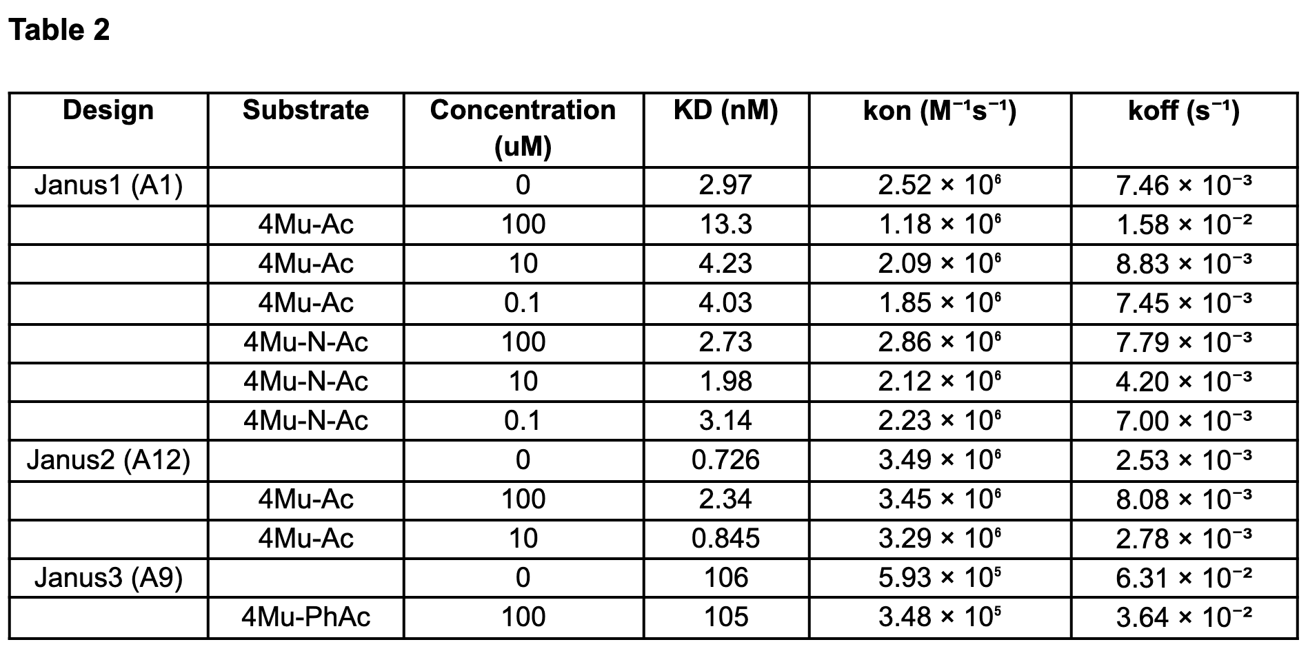
Substrate concentration vs K_D
Methods
Computational design of allosteric enzymes
Partial RFDiffusion for inactive state generation
de novo serine hydrolase enzymes from Lauko et al. as our starting active enzyme backbone (15)
RFDiffusion for effector generation
The resultant effectors were designed to be either allosteric activators or inhibitors depending on whether they were generated in the context of the active enzyme state or inactive enzyme state, respectively (15)